Call to Schedule Free Consultation at Over 45 Centers Worldwide!
Autoimmune
Cardiovascular
Endocrine
Gastrointestinal
Liver
Musculoskeletal
Neurological
Other
Respiratory/Pulmonary
Reproductive
Renal/Kidney
Urologic
Autoimmune
Cardiovascular
Endocrine
Gastrointestinal
Liver
Musculoskeletal
Neurological
Other
Respiratory/Pulmonary
Reproductive
Renal/Kidney
Urologic
DISEASE AWARENESS PAGE FOR R3 STEM CELL – FRACTURES
A fracture happens when there is a break in the continuity of a bone. Bones are the body’s supporting tissues, and they are continuous throughout their structure. A break along the body of any bone is called a fracture, and depending on the cause, the pattern may vary. But regardless of the cause, most fractures cause severe pain to the individual.
Fractures often heal over time with some medical treatment. The kind of treatment and attention given to a fracture depends on the cause, the extent of the fracture, as well as the complications that may arise. Some fractures can even heal without medical attention, although there are chances that the limb will be permanently deformed.
Fractures in the upper limb usually heal within 8 to 12 weeks. Lower limb fractures take longer, usually between 12 to 16 weeks. Some other factors determine the speed of healing. For example, the bones of children heal faster than those of adults.
Bones are connective tissues in the body, and they are made up of tiny bone cells (or osteo-cells). There are different kinds of bone cells, and they are all differentiated based on their function. While osteophytes make up the structure of the bone, osteoclasts are responsible for reabsorbing and remodelling the bone.
Any break in the continuity of a bone is a fracture. However, since bones connect at joints, a dislocation, in a sense, is also a break in continuity. The difference is that while fractures apply to a break along the shaft (main body) or end of a bone, dislocations apply to joints. This means that dislocation is the separation of two or more bones at their joints.
Dislocations are equally painful as fractures, and they can be just as deforming if not properly treated. Sometimes, it’s difficult to tell a fracture from a dislocation, especially when the fracture occurs very close to the joint end of the bone. In other cases, a fracture and a dislocation happen at the same time, complicating the condition. X-rays are used to tell these conditions apart from each other.
As mentioned earlier, there are different types of fractures, and they all have slightly different patterns. Fractures can be classified based on the pattern of the break on the bone, based on the cause, and based on the extent of damage to the bone and surrounding tissue.
Classified based on the extent of damage, fractures can be classified into:
A fracture is said to be open when one or more of the bone fragments break the skin and are exposed. This is commonly seen in traumatic fractures where the impact causes the bone to protrude out of the skin. Besides the difficulty in realigning the bony pieces, open fractures can have other complications like infections and slowed healing.
A closed fracture is one in which the broken bone remains within the body. That means that there’s no bone sticking out of the body. These kinds of fractures are considered to be less painful and easier to treat than open fractures. They also have fewer complications.
Traumatic fractures are the most common types of fractures, and they are caused by a severe trauma to the bone in question. Because bones are incredibly strong, the force required to cause a fracture is always significant.
A stress fracture occurs when a bone bears excessive load over time. This is often seen in the elderly. As mentioned earlier, bones are incredibly strong connective tissues, and often require great force to cause a fracture. However, in older people, conditions like osteoporosis weaken the bones, making it easier to sustain a fracture.
A pathological fracture is caused by disease conditions in the body. These are the most troublesome kinds of fractures because until the underlying condition is resolved, the individual is predisposed to sustaining future fractures. A condition that can cause recurrent fractures is Osteogenesis Imperfecta, commonly known as brittle bone disease.
As mentioned earlier, fractures can be caused by a couple of different factors. These factors are often involved in the way the fracture is named. The major cause of fractures is trauma, and it is very common in car accidents and falls from great heights.
Other causes of fractures include:
The prevalence of fractures varies among younger and older people, and among men and women. In men between the ages of 18 and 49, the incidence of fracture is about 94.8 in 10,000 people or roughly 1%. In women of the same age range, the incidence was 54.3 in 10,000 women or roughly 0.5% In men over the age of 50; the incidence is about 0.7% in men and 1.5% in women.
Older people have a bigger chance of sustaining fractures than younger people. This is because as we age, our bones lose density, and they get more brittle. Also, all the other supporting systems in the body like the joints and the muscles age and lose their strength and elasticity. All of this contributes to the increased likely hood of sustaining fractures.
People who are in high-stress jobs are at risk of getting stress fractures. This is because the repeated impact of the heavy loads on the bones weakens them over time.
Women are more predisposed to sustaining fractures than men. This is because women have lesser bone density and strength than men. Also, women lose more bone density as they grow older, compared to men.
Smoking puts a person at risk for sustaining a fracture because of its influences on hormones. Women who smoke have been shown to go through menopause earlier than women who don’t. It also leads to bone loss.
Symptoms of fractures are quite easy to identify. The most significant of them is an obvious deformity in the affected area. Any break in the continuity at the arms or legs can be easily seen. Other symptoms of fractures include
A fracture can be diagnosed by assessment alone, depending on the area. A fracture of the arm is easily identified. However, further tests like X-rays may be conducted to determine the extent and kind of fracture. It may also be conducted to rule out other suspicious. In some cases, MRIs may also be requested to determine the extent of damage to the surrounding tissues.
However, an X-ray is often all that’s necessary.
The treatment of a fracture depends on the severity of the fracture. Most fracture treatments follow these steps
The most important part of treating a fracture is reducing it. If it is a simple closed fracture, traction may be all that’s necessary. In traction, a force is applied to the distal end of the bone, and that moves it back into place
In other situations, surgery may be needed to reduce the fracture. A plate and screw may then be sued to hold the bones in place
After reduction, the bone pieces are held together, and the affected part is immobilized to allow the bone rest and heal. This may be achieved by the cast alone. In other situations, like an upper limb fracture, an arm sling may be necessary.
Some painkillers may be prescribed to reduce the pain associated with the fracture
Here are answers to many of the frequently asked questions we receive.
Arthritis can be classified into several types. Osteoarthritis is caused by the normal aging process of the body, or could be a result of injury.
Rheumatoid arthritis, the most common type of arthritis, happens due to autoimmune problems, wherein the body’s own immune system attacks healthy cells of the body. Infectious arthritis is caused by an infection to the joint. Patients with psoriasis may be affected by Psoriatic arthritis. Finally, excessive uric acid in the body can cause Gout, an arthritis that often starts at the toe.
Rheumatoid arthritis is an inflammatory form of arthritis. This disease causes chronic inflammation of the joints and can lead to deterioration of a variety of systems in the body. Rheumatoid arthritis occurs when the immune system attacks the lining of the membrane around the joints known as the synovium, which causes inflammation that thickens the synovium and ultimately deforms the cartilage and bone of the joints.
How common is it and who is at risk?
An estimated one percent of the entire world population suffers from rheumatoid arthritis. As the case with osteoarthritis, women are more likely to develop rheumatoid arthritis. Other groups at risk are people middle-aged or older as well as those suffering from obesity. Rheumatoid arthritis risk is inheritable, which may be accelerated by smoking or unknown environmental exposures.
Signs of the disease include swollen, tender joints that may feel stiff, particularly in the mornings. Fever, fatigue, and loss of activity are also common symptoms. Rheumatoid arthritis tends to manifest initially in the smaller joints like those in the fingers and toes. Symptoms are exhibited in joints of other body parts as the disease spreads.
Diagnosis begins with a physical test checking for redness, warmth, and swelling as well as the state of reflexes in the affected joints. Multiple blood tests are conducted as people with rheumatoid arthritis usually have elevated erythrocyte sedimentation rates and C-reactive protein levels. Additionally, X-rays, MRIs, and ultrasounds are conducted to track the spread of the disease.
While no cure exists as of now, research has revealed that the symptoms may be controlled by early application of disease modifying anti-rheumatic drugs (DMARDs). Steroids or nonsteroidal anti-inflammatory drugs may be prescribed depending on the case. Exercises to retain joint flexibility also provide some relief. If medications fail to slow down the disease, doctors may recommend surgery to repair or replace joints or swollen tendons.
Juvenile Idiopathic Arthritis (JIA) is the most prevalent form of arthritis within kids. It is characterized by persistent pain, swelling, and stiffness in the joints. The disease occurs when the body’s own tissues are attacked by its cells. In some cases, it can create more serious complications such as growth problems and eye inflammation.
It is the most common form of arthritis diagnosed in children under the age of sixteen. Certain gene mutations can make the body more vulnerable to the external environment, which can result in Juvenile Idiopathic Arthritis occurring. Although reasons are unknown, girls are more susceptible to Juvenile Idiopathic Arthritis than boys.
Commonplace symptoms include joint pain or a limp caused by the pain, swelling of joints and stiffness of the joints that results in clumsiness by the child. In some cases, the disease can manifest as high fever and rashes.
Diagnosis for Juvenile Idiopathic Arthritis is difficult as the pain and swelling in the joints could be attributed to a number of diseases. Quite a few blood tests are conducted to determine the status of markers like erythrocyte sedimentation rate, C-reactive protein levels, and quantity of anti-nuclear bodies, among others. However, many children with Juvenile Idiopathic Arthritis show no abnormalities in these tests.
The treatment for Juvenile Idiopathic Arthritis is geared towards making sure the child can maintain a regular level of physical activity. This can include different types of medication like nonsteroidal anti-inflammatory drugs to reduce pain and swelling or disease modifying anti-rheumatic drugs in more severe cases. Biologic agents may be recommended to help prevent joint damage. Physical therapy can also help the child maintain a full range of motion with or without joint supports or splints based on the situation. Surgery is the most extreme option used to improve the position of the affected joint.
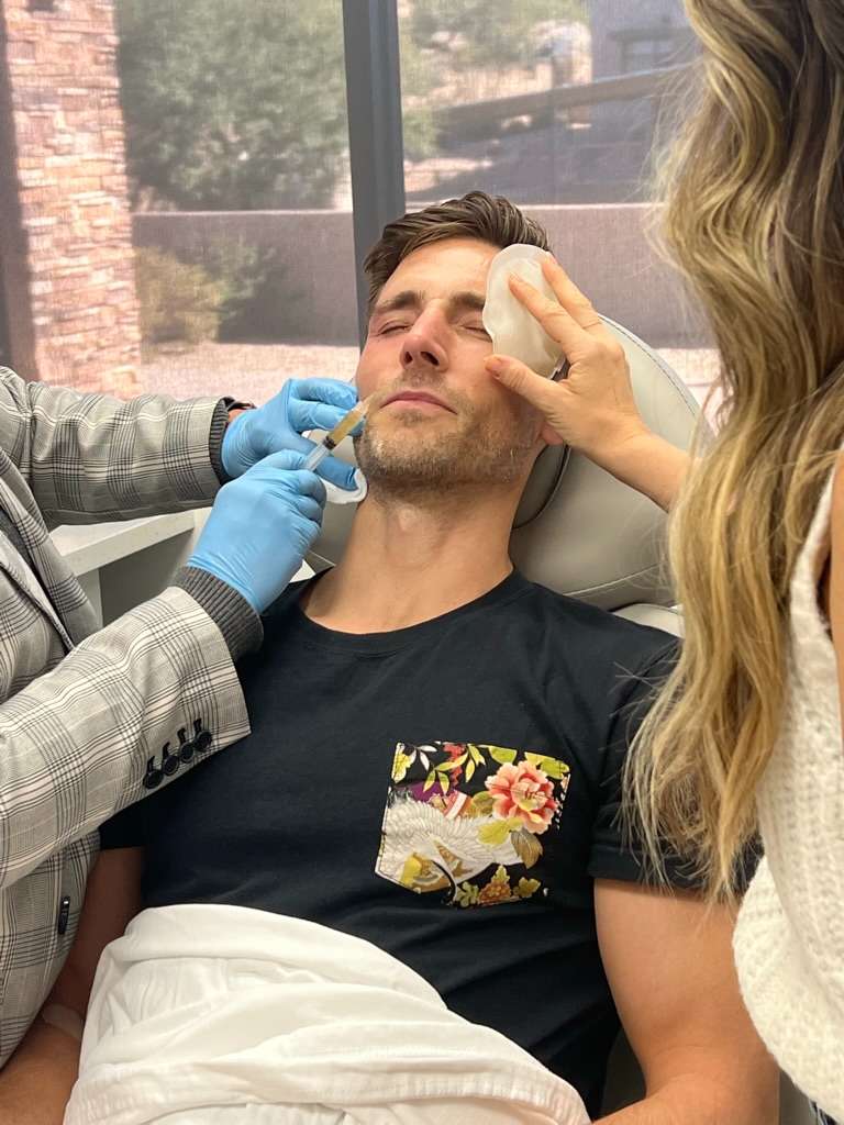
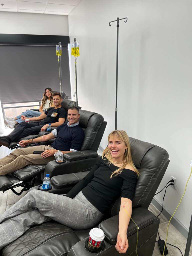
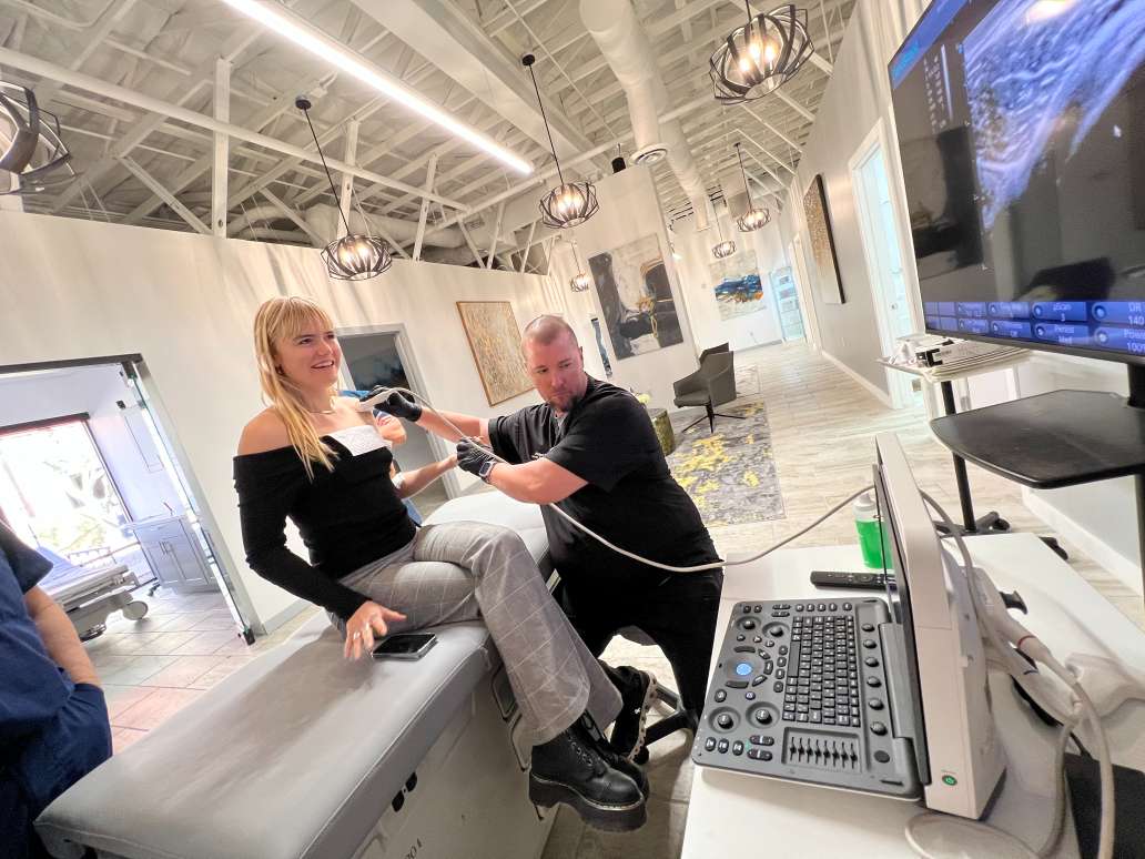
Brand Ambassador Gallery
The USA stem cell leader offers procedures in
7 Countries including:
SUCCESS STORIES
R3 STEM CELL MASTER CLASS
Learn everything you need to know about the ever expanding field of regenerative medicine in this 8 part series that includes over four hours of entertaining content!
R3 STEM CELL INTERNATIONAL
R3 Stem Cell International includes 45 clinics in 7 countries. These Centers of Excellence treat all types of conditions with safe, effective protocols by expert stem cell physicians.
FREE STEM CELL CONSULTATION
R3 Stem Cell offers a no cost consultation to see if you or a loved one is a candidate for regenerative cell therapies including cytokines, growth factors, exosomes, and stem cells.
PROVIDER PARTNERSHIP
The R3 Partnership Program offers providers an all-in-one regenerative practice program including marketing, consultations and booked procedures!
The information provided by R3 Stem Cell is not a substitute for professional medical advice, diagnosis, or treatment. Individual results may vary and only your medical professional can explain all the risks and potential benefits of any therapy based on your circumstances. R3 Stem Cell does not recommend or endorse any specific tests, products, procedures, opinions, or other information that may be mentioned on this website. Reliance on any information provided by R3 Stem Cell, its employees, others appearing on this website at the invitation of R3 Stem Cell, or other visitors to the website is solely at your own risk. R3 Stem Cell is not responsible for the outcome of your procedure. The FDA considers stem cell therapy experimental at this point.
CALIFORNIA
FLORIDA
GEORGIA
HAWAII
IDAHO
ILLINOIS
INDIANA
IOWA
KANSAS
KENTUCKY
LOUISIANA
MARYLAND
MASSACHUSETTS
MICHIGAN
MINNESOTA
MISSISSIPPI
MISSOURI
NEBRASKA
NEW JERSEY
NEW YORK
NEW MEXICO
NEVADA
NORTH CAROLINA
OHIO
OKLAHOMA
OREGON
PENNSYLVANIA
RHODE ISLAND
SOUTH CAROLINA
SOUTH DAKOTA
TENNESSEE
The USA stem cell leader offers procedures in
7 Countries including:
Copyright © 2017-2024 R3 Stem Cell. All Rights Reserved.