
Amniotic membrane and its Use in Tendon Repair
Introduction
A multilayer tissue, amniotic membrane forms the innermost layer of the amniotic sac which surrounds the developing fetus. It basically comprises of 5 layers: A single layer of epithelial cells, a thick basement membrane, compact layer, layer of fibroblast and a spongy layer that is adherent to the chorion. Amniotic membrane has several functions which include the synthesis of cytokines and growth factors, regulation of pH, transport of solutes and water, and acting as a permeable barrier to amniotic macromolecules.1
Advantages of using Amniotic membrane therapy
The advantage of Amniotic tissue is that it is readily available, as it is usually discarded after birth. As this tissue does not pose any harm or danger to the fetus and the mother, there are no ethical concerns associated with using and obtaining embryonic stem cells. Amniotic membrane is composed of an extracellular matrix, that acts as a scaffold for structural support for cells and cellular attachment along with attachment for collagen types I, III, IV, V and VI, growth factors and hyaluronic acid2. In addition to this it also possess antimicrobial properties due to the presence of Beta-defensins and anti-inflammatory effects by inhibiting the inflammatory cascade.
Amniotic membrane has been in used for the past 100 years to treat burns, ocular diseases and chronic wound with favorable outcomes and without any adverse effects. Amniotic cells do not possess any type of tumorigenicity unlike the embryonic stem cells which may be tumorigenic. This lack of tumorigenicity and any type of immune response is due to the fact that the amniotic cells do not express the human leukocyte class II antigen and express only class I antigens, that too in small amounts.3
Indications
Amnion applications in orthopedic surgery may be numerous, but development is ongoing4. Given the vast array of in vitro and in vivo animal data supporting the benefits of amnion in tissue regeneration, orthopedic surgeons and researchers should place emphasis on conducting clinical studies to validate the safety and efficacy of amniotic cells in the treatment of orthopedic conditions.
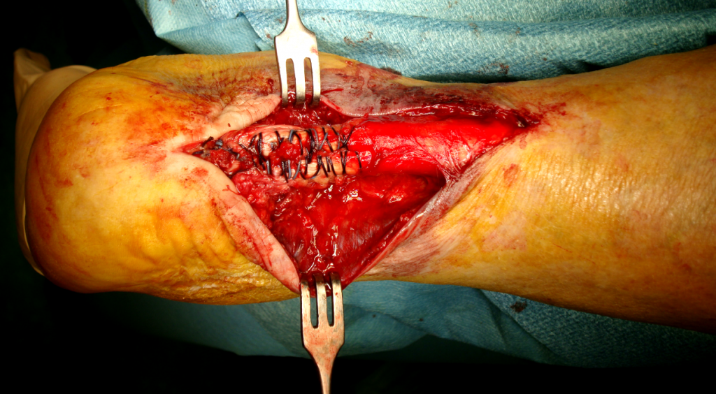
The use of amniotic membrane in orthopedic surgery is increasing. Tendons, cartilages and ligaments are virtually avascular structures, this means that they have no blood supply to provide them with nutrients and building blocks to rebuild and repair any type of damage inflicted to them. This is the reason why tendon repair is generally a lengthy process. With the use of amnion-derived therapy, physicians can now introduce a high concentration of proteins, cytokines, growth factors and essential nutrients to hasten the process of healing.
A study conducted in 2009 by Fahir Demirkan et all on the use of amniotic membrane for the repair of flexor tendon demonstrated that with the use amniotic membrane for repair, the healing process increased considerably and the tendon healed without formation of any adhesions.5 A study conducted in 2012 by Barboni and colleagues used amniotic epithelial cells to be implanted into an artificially created Achilles tendon of a sheep, inducing effective structural and mechanical recovery in the detects at a rate which was faster than in the control group. This therapy induced a centripetal healing process at first around the healthy tissue of the tendon and then progressed towards the core of the tendon. This is highly suggestive of recruitment of native progenitor cells towards the defect and generation of a new tendon matrix.6
In a study published in 2013 by Justin Philip et all, use of amniotic membrane was assessed in the repair of the Achilles tendon in a model of rats. The tendons of the rats were excised and then repaired. A control group was established which did not receive the amniotic membrane therapy. After 4 weeks the groups were assessed for the tensile strength, Young’s modulus and the breaking strength of the tendon. Mechanical testing showed that the group treated with the amniotic membrane had a higher Young’s modulus and high tensile and breaking strength.7
Another study by Kueckelhaus and colleagues investigating the role of cytokines derived from amnion in healing the Achilles tendons of rats, reported an improvement in the mechanical properties of healing tendons as compared to the control group.8
Amniotic membrane therapy is a safe treatment without having any tumorigenic effects, producing essential growth factors that have shown benefits as aids in regeneration for soft tissues and human bones. Amnion applications for the use in orthopedic surgery are numerous, but this is a developing field. Most of the research present in this field is done on animal with only a few cases being reported on humans. As the amniotic membrane does not differ in an animal or a human, use of amniotic membrane therapy is safe and could yield promising benefits in the field of orthopedic surgery as seen with in vitro and in vivo testing. This should be explored and brought into light.
References
- Mamede AC, Carvalho MJ, Abrantes AM, Laranjo M, Maia CJ, Botelho MF. Amniotic membrane: from structure and functions to clinical applications. Cell and tissue research. 2012 Aug 1;349(2):447-58.
- Gupta A, Kedige SD, Jain K. Amnion and chorion membranes: potential stem cell reservoir with wide applications in periodontics. International journal of biomaterials. 2015 Dec 6;2015.
- Miki T, Strom SC. Amnion-derived pluripotent/multipotent stem cells. Stem Cell Reviews and Reports. 2006 Jun 1;2(2):133-41.
- McIntyre JA, Jones IA, Danilkovich A, Vangsness Jr CT. The Placenta: Applications in Orthopaedic Sports Medicine. The American Journal of Sports Medicine. 2017 Apr 1:0363546517697682.
- Demirkan F, Colakoglu N, Herek O, Erkula G. The use of amniotic membrane in flexor tendon repair: an experimental model. Archives of orthopaedic and trauma surgery. 2002 Sep 1;122(7):396-9.
- Barboni B, Russo V, Curini V, Mauro A, Martelli A, Muttini A, Bernabò N, Valbonetti L, Marchisio M, Di Giacinto O, Berardinelli P. Achilles tendon regeneration can be improved by amniotic epithelial cell allotransplantation. Cell transplantation. 2012 Nov 1;21(11):2377-95.
- Philip J, Hackl F, Canseco JA, Kamel RA, Kiwanuka E, Diaz-Siso JR, Caterson EJ, Junker JP, Eriksson E. Amnion-derived multipotent progenitor cells improve Achilles tendon repair in rats. Eplasty. 2013;13.
- Kueckelhaus M, Philip J, Kamel RA, Canseco JA, Hackl F, Kiwanuka E, Kim MJ, Wilkie R, Caterson EJ, Junker JP, Eriksson E. Sustained release of amnion-derived cellular cytokine solution facilitates Achilles tendon healing in rats. Eplasty. 2014;14.