
Amniotic Membrane and Its use in Periodontics
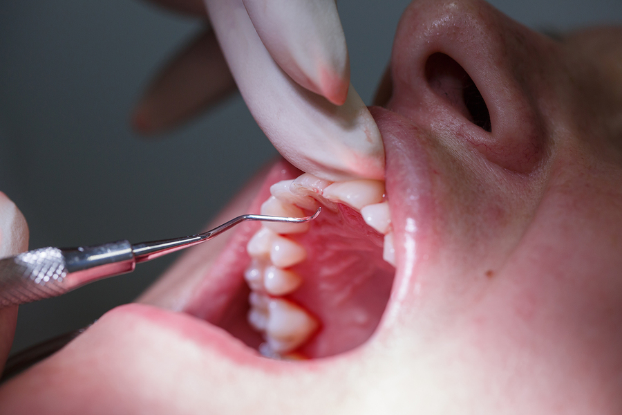
Introduction
Amniotic membrane surrounds the fetus and the amniotic fluid. It is a highly flexible membrane which is why it can be easily separated from the chorion. It contains of two cell types; the epithelial cells that are derived from the embryonic ectoderm and the amnion mesenchymal cells that are derived from the embryonic mesoderm.
At microscopic level it is a thin, avascular and transparent composite membrane that is made up of three major layers; a single epithelial layer, a thick basement membrane and the mesenchymal layer consisting of mainly collagen types I, III, IV, V and VI1.
How does the amniotic membrane help?
The extracellular matrix is made up of substances that give the amniotic membrane its structural integrity and it contains a variety of specialized proteins which include proteoglycans, glycosaminoglycans, fibronectin, laminins and other materials similar to these.
The basal lamina contains proteoglycans like heparin sulfate which is also the major proteoglycans found in the Gingiva. The spongy layer of the amniotic membrane contains an abundance of hydrated proteoglycans and glycoproteins; they form a non-fibrillar network alongside collagen2.
The matrix of the amniotic membrane has an abundance of growth factors like basic fibroblast growth factor, transforming growth factor beta (TGF-B), keratinocyte growth factor, nidogen growth factor and epidermal derived growth factor which promotes periodontal regeneration. These growth factors provide a nurturing environment that accelerates healing and mimic the stem cells for ex vivo growth3.
Properties of Amniotic Membrane
Amniotic membrane possesses multiple beneficial properties that make its use in dentistry and periodontic revolutionary. It has the following properties:
- Epithelialization
- Anti-inflammatory
- Anti-viral and anti-microbial
- Anti-Scarring
- Angiogenesis (formation of new blood vessels)
- Immunomodulatory properties
There has been an increase in the use of the amniotic membrane clinically as an allograft material for the management of chronic and acute wounds, for reducing scar tissues, as a soft tissue regeneration graft and as a barrier membrane4. It is easily available and it is cost effective to preserve.
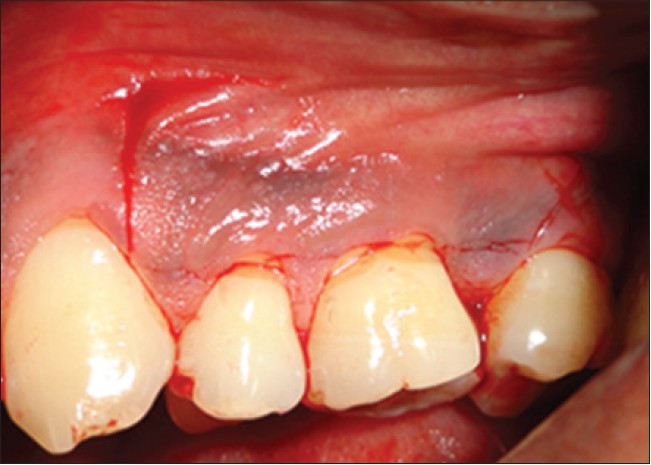
Amnion allograft might be an effective and suitable alternative to the connective tissue graft that is used in procedures to cover the denuded root surfaces and reduce recession depth5. The graft is also a reliable method to cover the exposed periosteum as it serves as an alternative to mucosal and skin grafts6.
What are its indications in Periodontics?
Amniotic membrane has numerous applications in various fields of medicine, including burn management, reconstruction of the oral cavity, vagina and the bladder, arthroplasty, tympanoplasty and ophthalmology to name a few. Recently amniotic membrane has found application in the field of periodontics.
Recent literature being published indicates that amniotic membrane provides positive results in terms of increased tissue thickness, root coverage, increased attached gingival tissue, as a barrier membrane for intrabony defects, healing of soft and hard tissue and provides excellent esthetic results in term of color and texture match7,8.
As the membrane is considerably thin, along with having a self adherent nature, it intimately adapts to the contours around the roots and defects, eliminating the need to use sutures. This makes the procedure less technical and decreases the surgical time significantly.
A study conducted in 2010 by Velez et all evaluated amniotic membrane for tissue healing in terms of epithelialization, lesion size, infection, pain, inflammation and scarring in dental implant surgery. Their study revealed that the cryopreserved amniotic membrane was beneficial and effective in helping wound healing, cicatrisation, reinforced adhesion, supported growth of epithelium and decreases the pain level in their subjects7.
A retrospective observation study conducted in 2013 by Holtzclaw D.J et all documented the use of amniotic membrane for guided tissue regeneration treatment for periodontal intrabony defects. Their observations revealed improved density of the bone fill as compared to the base line level. There are numerous benefits of using amniotic membrane over convention guided tissue regeneration membrane9.
Amniotic membrane has provided an alternative to conventional autograft tissue treatment for root coverage procedures. Tsuno et all have reported of successfully treating intraoral alveolar wounds during vestibuloplasty of a reconstructed mandible using a dry amniotic membrane10.
A histological study also demonstrated that use of amniotic membrane for gingival wound healing induces rapid epithelialization and formation of both collagen and granulation tissue while suppressing inflammation11.
Human amniotic membrane is a revolutionary material to be used as an allograft in wound management and in various fields of tissue medicine, engineering, regeneration biology and stem cell research. The clinical use of this membrane not only maintains the anatomical and structural integrity of the regenerated tissue but it also aids in hastening healing and reducing post operative scarring, providing a rich source of stem cells.
To conclude, the use of amniotic membrane in regenerative medicine is a revolutionary and innovative step towards treating numerous condition previously treated poorly.
References
- . Chopra A, Thomas BS. Amniotic membrane: A novel material for regeneration and repair. Biomimetics Biomaterials and Tissue Engineering. 2013;18(1):1-8.
- Parry S, Strauss JF. Premature rupture of the fetal membranes. New England Journal of Medicine. 1998 Mar 5;338(10):663-70.
- Fetterolf DE, Snyder RJ. Scientific and clinical support for the use of dehydrated amniotic membrane in wound management. Wounds: a compendium of clinical research and practice. 2012 Oct;24(10):299-307.
- Sharma M, Kotwal B, Mahajan N, Kharyal S. Amniotic Membrane in Periodontics: An Insight.
- Ghahroudi AA, Khorsand A, Rokn AR, Sabounchi SS, Shayesteh YS, Soolari A. Comparison of amnion allograft with connective tissue graft for root coverage procedures: a double-blind, randomized, controlled clinical trial. Journal of the International Academy of Periodontology. 2013 Oct;15(4):101-12.
- Yang S, Leong KF, Du Z, Chua CK. The design of scaffolds for use in tissue engineering. Part I. Traditional factors. Tissue engineering. 2001 Dec 1;7(6):679-89.
- Velez, W. B. Parker, M. A. Siegel, M. Hernandez, Cryopreserved Amniotic Membrane for Modulation of Periodontal Soft Tissue Healing: A Pilot Study, J. Periodontol, 81,1797- 1804 (2010)
- Rosen. A Case Report on Combination Therapy Using a Composite Allograft Containing Mesenchymal Cells With an Amnion–Chorion Barrier to Treat a Mandibular Class III Furcation, Clin. Adv. Perio, 3, 64-69 (2013).
- Holtzclaw DJ, Toscano NJ. Amnion–chorion allograft barrier used for guided tissue regeneration treatment of periodontal intrabony defects: A retrospective observational report. Clinical Advances in Periodontics. 2013 Jul 22.
- Tsuno H, Arai N, Sakai C, Okabe M, Koike C, Yoshida T, Nikaido T, Noguchi M. Intraoral application of hyperdry amniotic membrane to surgically exposed bone surface. Oral surgery, oral medicine, oral pathology and oral radiology. 2014 Feb 28;117(2):e83-7.
- Rinastiti M, Santoso AL, Sosroseno W. Histological evaluation of rabbit gingival wound healing transplanted with human amniotic membrane. International journal of oral and maxillofacial surgery. 2006 Mar 31;35(3):247-51.